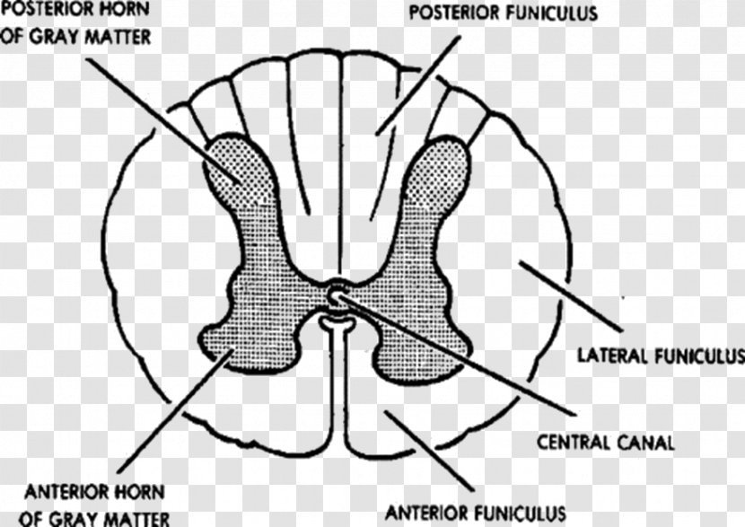
A cross-section of the anterior horn of gray matter, which is a type of vertebrae. labeled with the names of the bones and their structures. The anterior horn is located in the center of the image and is surrounded by the central canal. The central canal is located on the right side of the diagram. The anterior horn has a large, elongated bone in the middle, which appears to be a vertebral column. The bones are arranged in a way that they form a symmetrical pattern around the central bone. The bone is connected to the lateral funicularus, which extends from the central part of the body to the outer part of its head. There are also several lines connecting the bones, which are labeled with their names. These lines represent the different parts of the bone, such as the anterior and lateral functions. The lines are labeled as "Posterior Funicularus" and "Central Canal."
Overall, the image shows the anatomy of the posterior horn and its structures, which can be seen in a detailed and intricate manner.
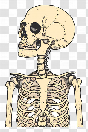
User juveeil uploaded the image
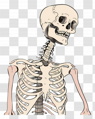
User juveeil uploaded the image
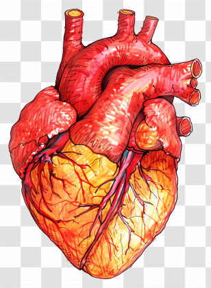
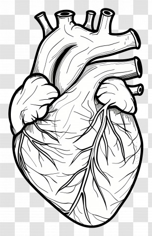
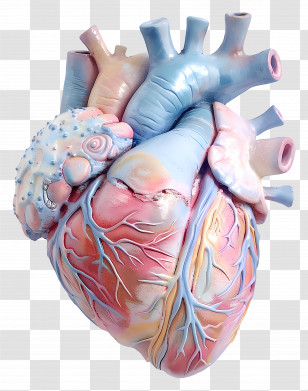
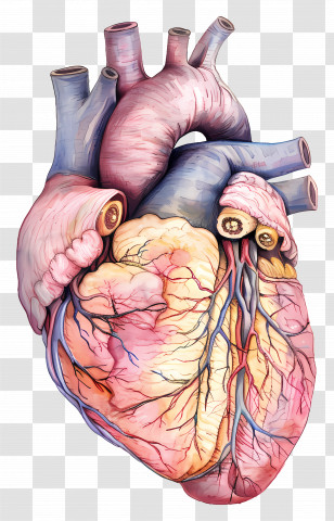
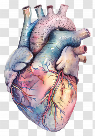
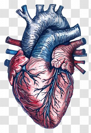
Spinal Cord Grey Matter Vertebral Column Central Nervous System Anatomy - Heart - Silhouette PNG
. The resolution of this PNG file is 880 x 623 pixels and it has a file size of 102.33 KB.Spinal Cord Grey Matter Vertebral Column Central Nervous System Anatomy - Heart - Silhouette PNG
You might also like these images below...